Skin Cancers: The Diagnostic Process
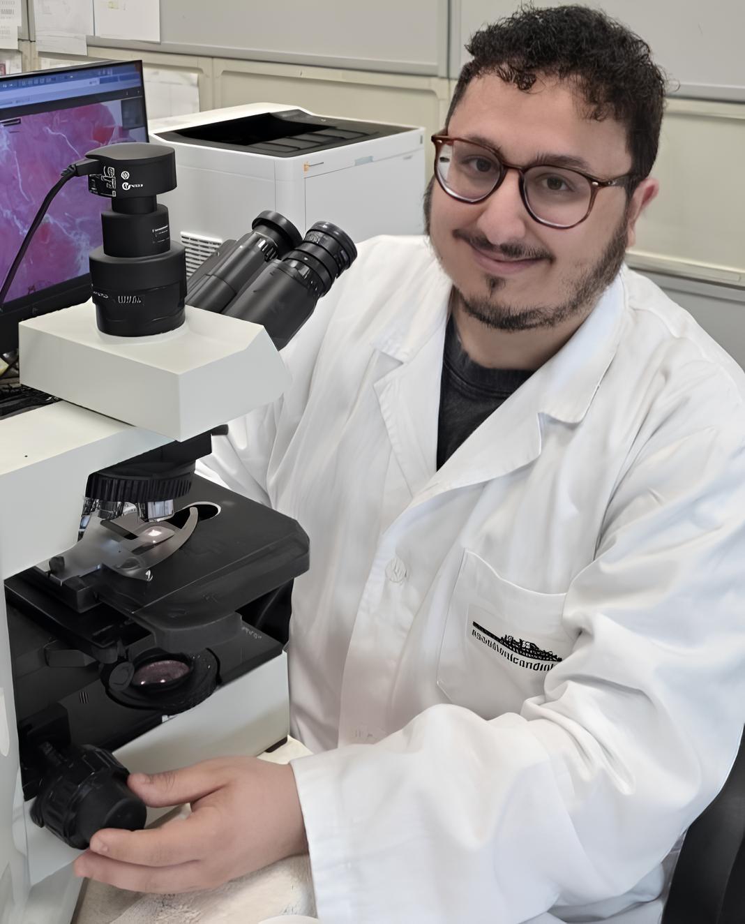
Dr.
Damian Carrot
Pathologist in the Division of Pathology
I skin cancers These include various types of tumors, each with different characteristics and levels of aggressiveness. In this article, we’ll look at how to recognize the symptoms, how a skin biopsy works, and what happens to the sample in the lab. Finally, we’ll explain how to interpret a histology report to determine which tests to order and how to develop the best treatment plan. Here’s everything you need to know.
Go to profile
What are the main types of malignant skin cancers, and how are they diagnosed?
There are several types of malignant skin cancers, each with distinct biological behavior. The most common is basal cell carcinoma, a tumor that primarily develops on skin that has been chronically and heavily exposed to sunlight and which, depending on the subtype, may grow more or less rapidly, with a tendency toward local invasiveness but a low likelihood of metastasis. Next among the most common are squamous cell carcinoma—which is more aggressive and tends to metastasize to lymph nodes—and the large family of melanomas, often aggressive tumors that can arise on skin subjected to high and prolonged sun exposure, on unexposed skin, in the eye, and in the mucous membranes.
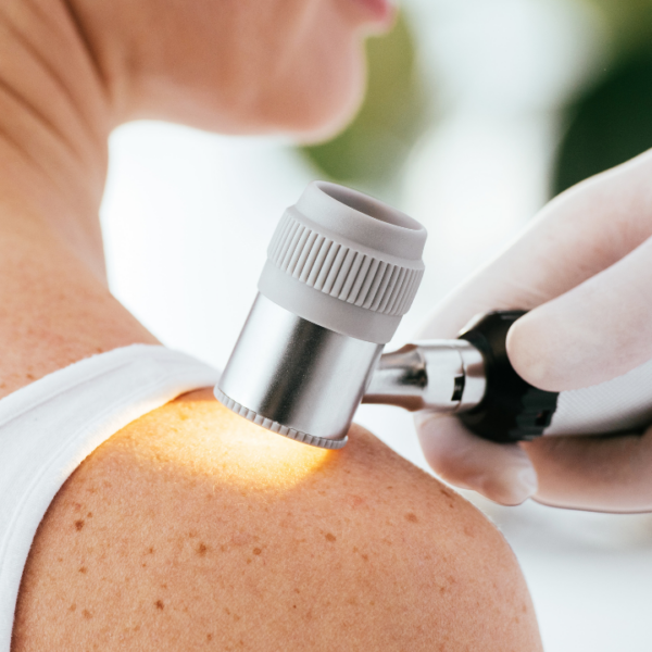
The diagnosis of skin cancers involves several professionals, starting with the patient, who performs periodic self-examinations of the skin and attends regular dermatological checkups. A surgeon (dermatologist, plastic surgeon, or general surgeon) may perform a biopsy on a suspicious lesion, and ultimately,the pathologist will make the definitive diagnosis after examining the sample in the laboratory and evaluating it under a microscope.
In certain cases, once a diagnosis has been made, various medical professionals—including an oncologist, a surgeon, a pathologist, a radiologist, and a radiation oncologist—meet to determine the most appropriate treatment plan for the patient.
What is a skin biopsy, and how is it performed?
Depending on the type of tumor, a skin biopsy involves the partial or complete removal of the lesion. In the first case, it is referred to as an“incisional”biopsy, performed solely for diagnostic confirmation; in the second case, it is referred to as an“excisional”biopsy, which involves the complete removal of the lesion.
A biopsy is necessary so that the pathologist can examine the nature of the lesion and determine all the parameters essential for the diagnosis and subsequent treatment plan. Sometimes it may be necessary for the patient to undergo a second procedure known as“radicalization”or“margin widening,” in which the surgeon removes the skin around the scar to ensure the most radical resection of the lesion possible.
What happens to the skin sample in the lab?
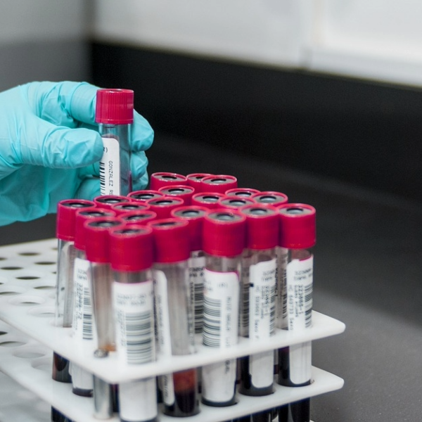
After the procedure, the specimen is immersed in a liquid called formalin, which preserves it in its original state, and then sent to the pathology department. There, it will undergo a series of steps—from sampling to processing—that will result in extremely thin tissue sections mounted on slides; once stained, the pathologist will be able to examine them under a light microscope.
The microscope allows for the observation of individual tumor cells, as well as their relationship to the surrounding tissues. The pathologist may also use special“immunohistochemical”stains to distinguish between different cell types. Sometimes the diagnosis can prove more complex than expected, so it is necessary to resort to molecular biology tests that analyze the tumor’s genetic profile and can guide the pathologist toward a specific diagnostic category.
What does the histopathology report say, and how do you interpret it?
A pathological report on a tumor consists of a detailed list of all the parameters and characteristics of the lesion necessary for the subsequent diagnostic and therapeutic process. Written in highly technical language,the report is intended for all medical professionalswho will be treating the patient; for this reason, when the patient receives it, it must be read and interpreted by the specialist who is treating them.
There is an extensive glossary of terms that may appear in a skin cancer report, including:
- resection margins: These indicate the quality of the lesion’s removal. If, under a microscope, the tumor extends to a certain distance from the margins—which are then defined as “clear” or “free”—further surgery to widen the resection may not be necessary; if, on the other hand, the tumor involves the resection margins, it means that part of it may not have been removed, making it necessary—when possible—to surgically expand the previous procedure. In specific cases, however, radiation therapy or drug therapies may be used;
- Breslow thickness: This refers to the thickness of the melanoma in millimeters —that is, how deep it has penetrated the skin—and is a key parameter that helps clinicians determine the tumor’s aggressiveness and stage. Above a certain threshold, it indicates the so-called “sentinel lymph node biopsy” should be performed;
- degree of differentiation: indicates how much the cancer cells “resemble” the tissue of origin; the more “well-differentiated” the tumor is, the less aggressive it tends to be;
- Vascular and perineural invasion: When the tumor spreads to blood and/or lymphatic vessels and to nerve structures, the risk of distant metastasis and local recurrence increases.
What additional tests might be ordered after the diagnosis?
The work of a pathologist does not always end with identifying the type of tumor and measuring its margins. Today, in fact, especially in the case of melanoma it is often necessary to study the underlying genetic alterations in order to identify the most effective treatment possible. The most classic example in melanoma is the assessment of the mutational status of the BRAF gene, which, in a specific molecular context, can allow the oncologist to use specific drugs that would otherwise be ineffective. The oncologist, in consultation with the pathologist and the molecular pathology laboratory, will request the molecular tests necessary for each specific case; these tests are typically performed on the tissue sample already surgically removed or, in certain cases, on a blood sample from the patient.