Basal Cell Carcinoma: What It Is and How It Is Treated
BY.
Dr.
Marco Rubatto
Division Representative for Skin Cancer Surgery Division
The basal cell carcinoma, also known as basal cell carcinoma, is a malignant skin tumor and the most common type of cancer in the world. Although this classification may sound alarming, it is a very slow-growing form of skin cancer that rarely spreads to other organs and, if diagnosed early, can be treated with excellent results. Here’s everything you need to know.
Go to profile
What is basal cell carcinoma, and how common is it?
Basal cell carcinoma is a malignant tumor that originates in the basal cells, the deepest layer of the epidermis. It is classified as malignant because it can invade nearby tissues and, if left untreated for a long time, cause significant local damage. However, unlike melanoma, it rarely metastasizes, which generally makes it less life-threatening.
It is the most commonly diagnosed skin cancer in the world: in Italy, the incidence is approximately 100 cases per 100,000 people (Source: Guidelines for Non-Melanoma Skin Cancers – Basal Cell Carcinoma). It primarily affects the areas of the body most exposed to the sun (face, nose, ears, scalp) and usually develops after age 50, although in recent years there has been an increase in cases among younger people as well.
Basal cell carcinoma is highly treatable if diagnosed early: the key is not to overlook skin lesions that do not heal or that change in appearance over time.
What are the risk factors, and at what stage of life does it develop?
The main risk factor for basal cell carcinoma is chronic exposure to UV rays, both from the sun and artificial sources. People who have spent many years outdoors for work or as part of their lifestyle, or those who have frequently used tanning beds, accumulate damage to their skin’s DNA over time, which can promote the development of the tumor.
A fair skin type (skin that burns easily, blond or red hair, light-colored eyes) is an additional risk factor.Immunosuppression, such as that associated with organ transplants or immunosuppressive therapies, also increases the risk, as does exposure to arsenic or ionizing radiation in occupational or medical settings.
Basal cell carcinoma most often develops after age 50, when the damage accumulated over a lifetime begins to manifest. However, sun exposure during one’s youth—and sunburns during childhood, in particular—is a key factor in determining future risk.
The sun is one of the main causes, but the damage accumulates silently over decades: protecting your skin when you’re young is an investment in your long-term health.
How does it manifest, and what are the initial symptoms?
The appearance of basal cell carcinoma is often misleading: it may look like a small sore that won’t heal, a scab that keeps coming back, or a small, shiny, translucent bump with rolled edges and visible blood vessels on the surface. It is precisely this resemblance to harmless lesions that causes it to be overlooked for months or even years.
The most commonly affected areas are the face (particularly the nose, cheeks, and forehead), the ears, the neck, and the scalp. There are several clinical variants: the nodular form is the most common and typically appears as a pinkish pearl; the superficial form, on the other hand, appears as a red, scaly patch; the sclerodermiform form, which is rarer but more insidious, has poorly defined edges and can be difficult to detect even to the trained eye.
A wound that hasn’t healed for more than three or four weeks in an area exposed to the sun should not be ignored: it is the most common sign of basal cell carcinoma.
What are the stages of diagnosing basal cell carcinoma?
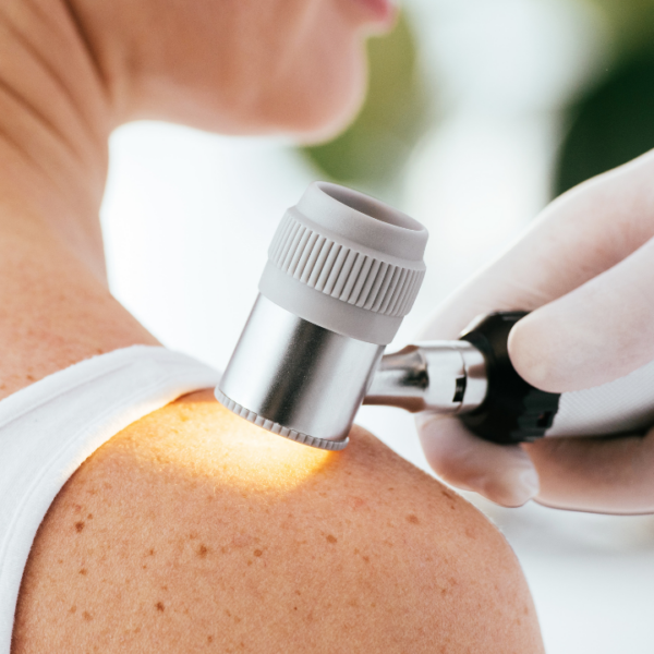
The diagnosis of basal cell carcinoma begins with a dermatological examination, during which the doctor examines the lesion and evaluates it using a dermoscope, an instrument that allows for a detailed analysis of the skin’s superficial structures. In many cases, this examination alone is sufficient to guide the diagnosis.
Definitive confirmation, however, always comesfrom a histological examination: a small sample of tissue is taken via biopsy and analyzed under a microscope by a pathologist, who identifies the type of tumor, its subtype, and the degree of invasion into the tissues. In selected cases—especially for large lesions or those in anatomically complex areas—further evaluation may be required using imaging tests such as ultrasound or magnetic resonance imaging (MRI).
A biopsy is nothing to be afraid of: it’s a small sample collection—often performed on an outpatient basis—that provides accurate and essential information for choosing the most appropriate treatment.
Can basal cell carcinoma be prevented?
Preventing basal cell carcinoma relies on simple but consistent habits. The most important is sun protection: using a sunscreen with an adequate SPF (Sun Protection Factor) every day—even in winter and in the city—reduces the accumulation of UV damage over time. It is essential to avoid sun exposure during the middle of the day (between 10 a.m. and 4 p.m.), wear hats and protective clothing, and avoid tanning beds.
Equally important isregular self-examination of the skin: checking your body regularly for new lesions or changes in existing ones allows you to detect any signs early on. This should be combined with dermatological checkups, which should be more frequent for those who have already had a basal cell carcinoma or have high risk factors.
Sunscreen is the most effective way to reduce the risk of developing the most common form of skin cancer in the world.
How is basal cell carcinoma treated?
The standard treatment for basal cell carcinoma is excisional surgery: the tumor is removed along with a margin of surrounding healthy tissue to ensure that it is completely removed. This procedure can generally be performed on an outpatient basis or in a day hospital under local anesthesia.
For lesions located in areas that are sensitive from an aesthetic or functional standpoint (such as the nose, eyelids, or lips), Mohs surgery is used—a technique that allows the tumor to be removed layer by layer, with the margins analyzed in real time to preserve as much healthy tissue as possible.
In cases where surgery is not an option or for small, superficial lesions, there are effective alternatives: photodynamic therapy, cryotherapy with liquid nitrogen, and laser treatment. For the very rare cases of advanced or inoperable basal cell carcinoma, targeted drugs are available that work by blocking the molecular mechanisms underlying tumor growth; when these prove insufficient, immunotherapy may be considered. After treatment, dermatological follow-up is essential: people who have had a basal cell carcinoma have an increased risk of developing a second one.