Melanoma: Prevention and Early Diagnosis
BY.
Dr.
Marco Rubatto
Division Representative for Skin Cancer Surgery Division
Melanoma is a type of skin cancer that often causes fear. Recognizing the symptoms of melanoma early is crucial: in fact, early diagnosis significantly increases the chances of recovery. In this article, we answer the most frequently asked questions about what melanoma is, who is at risk of developing it, and what to do to protect yourself.
Visit profile
What is melanoma, and how common is it in Italy?
The melanoma is a malignant skin tumor that originates in melanocytes, the cells responsible for producing melanin, the pigment that gives skin its color. Unlike other skin cancers, such as basal cell carcinoma or squamous cell carcinoma, melanoma is more likely to spread to other organs if not treated early.
In Italy, the numbers are on the rise: in 2024, there were an estimated 12,941 new cases of skin melanoma, with 7,069 in men and 5,872 in women (Source: AIOM – Cancer Statistics in Italy 2025). It can strike at any age, but it is particularly common among people aged 30 to 60, making it one of the most common cancers in young adults.
Detecting melanoma in its early stages radically changes the outlook: early diagnosis is the most important factor in the prognosis.
Who is most at risk of developing melanoma?
Certain factors significantly increase the likelihood of developing melanoma. The first is a fair skin type: people with fair skin, hair, and eyes are naturally less protected from UV rays and are therefore at greater risk. Added to this are sunburns—especially those sustained during childhood and adolescence—and the use of tanning beds, now recognized as a definite risk factor.
Family history also plays an important role: having a first-degree relative who has had melanoma increases an individual’s risk. The same applies to people who have a large number of moles or have had suspicious skin lesions in the past. In some cases, there is a genuine genetic predisposition linked to mutations in specific genes, such as CDKN2A or BRAF, which can be investigated at specialized centers.
Knowing your risk factors is the first step: those in higher-risk groups should start seeing a dermatologist sooner and have checkups more regularly.
Why is it important to use sunscreen to prevent melanoma?
UV rays (both UVA and UVB) can damage the DNA of skin cells, leading over time to mutations that may result in the development of cancer. Sunscreen is the most effective way to reduce this risk, provided it is used correctly.
A high SPF ( Sun Protection Factor)—50 or higher—is recommended especially for fair skin types, children, and anyone spending time outdoors during the middle of the day, when the sun’s rays are strongest. The sunscreen should be applied generously to all exposed areas and reapplied every two hours or after swimming. A common misconception is that a tan protects against the sun’s rays: in reality, it offers minimal protection, equivalent to an SPF of about 4.
Sunscreen isn’t just a cosmetic choice, but a concrete preventive measure: starting to use it as a child significantly reduces the risk over the course of a lifetime.
What is the ABCDE rule used for in melanoma?
The ABCDE rule is a practical tool that allows anyone to examine their moles and recognize any warning signs. The five letters correspond to five criteria to evaluate:
A for Asymmetry: A normal mole is symmetrical; if the two halves do not match, it’s a sign that warrants further investigation. B for Borders: The borders should be regular and well-defined; jagged or irregular borders are a red flag. C for Color: A healthy mole has a uniform color; the presence of multiple shades (brown, black, red, white) warrants attention. D for Diameter: Lesions larger than 6 mm warrant a medical evaluation, even though melanomas can be smaller. E for Evolution: Any change over time (in shape, color, size, or thickness) is the most important sign of all.
Self-examination is useful, but it has its limitations: the untrained eye may not notice subtle changes, and some areas of the body are difficult to observe. For this reason, it is never a substitute for a specialist examination.
Performing a self-exam of your skin is a simple habit that can make all the difference: if you notice anything that seems unusual, it’s best to see a dermatologist right away.
What are the main checks to perform, and when should they be done?
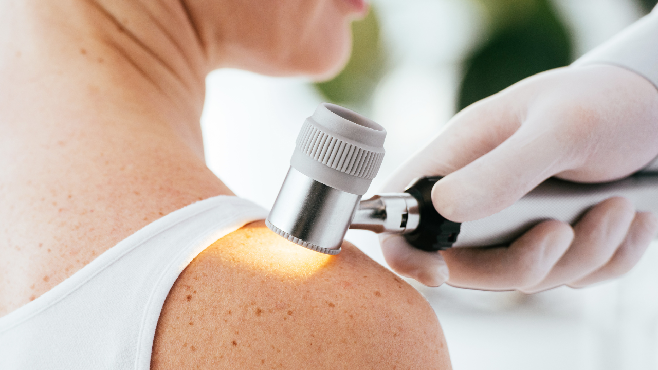
Having moles checked by a dermatologist is the most effective form of secondary prevention for melanoma. A dermatological exam involves the use of dermoscopy, a tool that allows skin lesions to be examined at high magnification, revealing details invisible to the naked eye. In some cases, a full mole mapping is also performed, which is useful for tracking any changes over time using standardized photographs.
The frequency of screenings depends on an individual’s risk profile: people with many moles, a family history of melanoma, or a history of suspicious lesions should have a screening every 6 to 12 months. For those without specific risk factors, an annual screening is still recommended. Your primary care physician can be your first point of contact to help determine when it is time to see a specialist.
It’s best not to wait until you notice something unusual before scheduling an appointment: routine checkups are designed precisely to detect problems that aren’t yet visible.
What are the main treatments for melanoma?
The treatment of melanoma depends largely on the stage at which it is detected. When diagnosed early, surgery is almost always curative: the melanoma is removed along with a safety margin of healthy tissue around it, in a procedure that is generally straightforward and performed on an outpatient basis.
In more advanced cases, where the tumor has spread to the lymph nodes or other organs, systemic therapies are used.Immunotherapy harnesses the patient’s immune system to fight cancer cells and is now the standard of care for many metastatic melanomas. Targeted therapy, on the other hand, is indicated for melanomas with specific genetic mutations (such as the BRAF gene mutation) and works by directly blocking the tumor’s growth mechanisms. After treatment, follow-up is essential: regular checkups and, in high-risk cases, diagnostic imaging tests allow for the early detection of any recurrences.